Aug 17, 2026
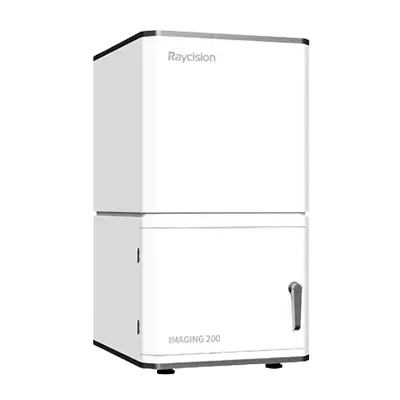
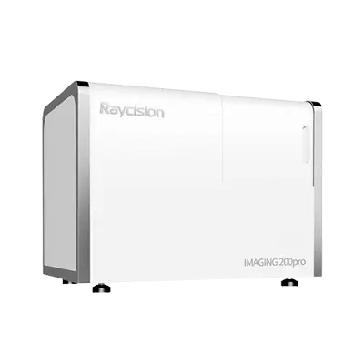
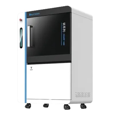
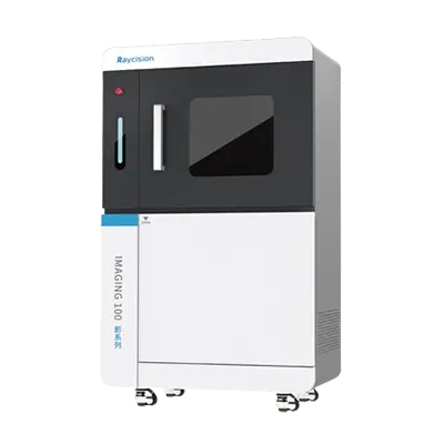
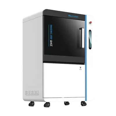
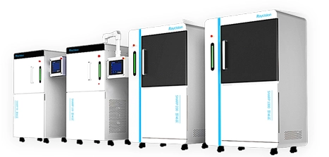
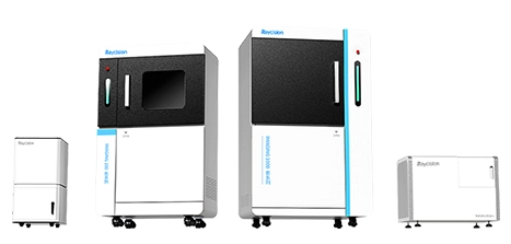
In preclinical radiation studies, a source that can deliver a selected dose is only the starting point. Researchers also need controlled dose rates, reproducible animal positioning, localized beam delivery, anatomical image guidance, treatment planning, and longitudinal response assessment. An X-ray radiation system can support a wide range of preclinical research, such as radiobiology, oncology, drug development, immunology, and combined-therapy studies. The appropriate configuration depends on whether a study requires whole-body irradiation, localized exposure, X-ray or CT guidance, conformal treatment, or molecular image-guided targeting. Raycision’s radiation portfolio includes the SHARP 100 X-ray Irradiator, SHARP 100pro X-Ray Image-Guided Radiation System, SHARP 200 CT-Guided Radiation System, and SHARP 1000 Multimodality Image Guided Precision Radiation System. These platforms provide different levels of imaging, targeting, dose calculation, and treatment-response evaluation for small animal research.
Unlike traditional gamma irradiators that rely on radioactive sources such as cobalt-60 or cesium-137, X-ray irradiators generate radiation electrically, allowing the source to be turned on for operation and switched off when treatment is complete. This approach avoids radioactive-source decay and eliminates the need to handle, store, or dispose of radioactive isotopes. X-ray irradiators also offer adjustable voltage, current, dose rate, beam size, and irradiation geometry. These controls support protocols ranging from broad-field exposure to localized irradiation of a tumor or selected anatomical region. Typical research applications include:
Total-body or partial-body irradiation
Tumor-growth and treatment-response studies
Normal-tissue radiation studies
Radiation sensitizer and protector evaluation
Combined radiotherapy and drug studies
Immunological and inflammatory research
Material and biological sample irradiation
The system should be selected according to the biological question being addressed. A straightforward irradiation study may not require integrated imaging, while anatomically precise tumor treatment needs image guidance and planning tools.
The first selection question is whether the study requires broad exposure or highly localized treatment. For routine irradiation protocols, the SHARP 100 provides adjustable dose rates from 0.01 to 100 Gy/min. Its listed dose uniformity is at least 95%, and adjustable beam collimators enable localized irradiation when a smaller treatment field is required. In addition, the system maintains an external dose rate below 0.5 μSv/h, so no additional shielding is required. Local installation requirements should nevertheless be reviewed according to applicable institutional and regional regulations. Researchers should define:
| Study requirement | Selection consideration |
|---|---|
| Exposure area | Whole body, organ region, tumor, or sample |
| Animal model | Mouse, rat, or another supported model |
| Dose range | Minimum, normal, and maximum protocol dose |
| Dose rate | Conventional, high-dose-rate, or ultra-high-dose-rate |
| Fractionation | Single exposure or repeated fractions |
| Positioning | Manual setup or image-verified targeting |
| Throughput | Number of animals or samples per session |
A suitable animal irradiator should cover the required protocols without forcing researchers to operate repeatedly at the limits of its dose-rate or field-size range.
An image-guided small animal irradiator may be appropriate for:
Orthotopic tumor models
Brain or lung targets
Bone and spinal studies
Organ-specific irradiation
Repeated fractionated treatment
Studies requiring improved setup reproducibility
Manual positioning may be sufficient for broad irradiation, but it introduces uncertainty when the target is small or located near radiosensitive tissue. Image guidance allows researchers to verify internal anatomy before radiation delivery rather than relying only on external landmarks. The SHARP 100pro adds high-resolution X-ray imaging to the irradiation workflow. It is designed to visualize and target anatomical regions in mice and rats while retaining adjustable dose rates, high dose uniformity, and collimator-based localized irradiation. Researchers should review image quality, positioning procedures, field verification, collimator options, and the time required to move from imaging to irradiation. These workflow factors influence both targeting accuracy and daily animal throughput.
Two-dimensional X-ray images can support positioning, but CT adds volumetric anatomical information. With CT, researchers can identify a target in three dimensions, contour surrounding structures, calculate dose distribution, and evaluate how beams interact with the complete anatomy. The SHARP 200 combines micro-CT imaging with image-guided radiation delivery. Its listed imaging resolution is no greater than 150 μm voxel size, and it includes GPU-accelerated reconstruction, adjustable collimators, target localization, conformal arcs intended to mimic clinical IMRT, and accelerated dose computation. The SHARP 200 also offers an ultra-high-dose-rate (UHDR) module with output up to 50 Gy/s, enabling UHDR-RT (FLASH-RT) studies. These studies require careful protocol design, dosimetry, validation, and interpretation because dose per pulse, total dose, field geometry, and biological endpoint can all affect experimental results. A CT image-guided radiation system is most valuable when researchers need to reproduce a clinical-style workflow involving imaging, contouring, planning, treatment delivery, and subsequent evaluation.
An anatomical image can show organs and structural boundaries, but some tumors or molecular targets may be difficult to identify by CT alone. Optical molecular imaging can provide functional information based on bioluminescent or fluorescent signals. The SHARP 1000 integrates X-ray, micro-CT, and optical molecular imaging for small animal radiation research. The platform uses multimodality guidance for target identification, treatment planning, radiation delivery, and outcome assessment. Its planning software incorporates GPU-accelerated Monte Carlo dose calculation, while registered multimodality images support quantitative evaluation after therapy. This workflow may benefit studies involving:
Small or poorly contrasted tumors
Metastatic disease models
Molecularly defined treatment targets
Longitudinal tumor-response assessment
Radiation combined with drugs or immunotherapy
Researchers should choose a multimodality system because it answers a defined biological question, not simply because it offers more imaging options.
Selecting an irradiator involves more than comparing dose rate and imaging resolution. Laboratories should review room layout, electrical requirements, animal preparation, anesthesia, monitoring, dosimetry, quality assurance, operator training, data storage, and maintenance support. Before selecting an irradiator, the following questions should be reviewed:
Which irradiation fields and collimators are available?
How are animals positioned and immobilized?
What dosimetry and calibration procedures are required?
Can the software support fractionated protocols?
What imaging dose is added during guidance?
How are plans, images, and irradiation records stored?
What training and preventive maintenance are provided?
A validated workflow should include output checks, field verification, positioning tests, and periodic quality assurance appropriate to the laboratory’s research program.
It delivers controlled radiation to animal models or biological samples for radiobiology, cancer research, drug development, immunology, and treatment-response studies.
No. An X-ray system generates radiation electrically, while a gamma irradiator uses a radioactive isotope. The two technologies differ in source management, energy characteristics, regulation, and maintenance.
Image guidance is valuable when radiation must be delivered to a small or internal target while limiting exposure to surrounding tissues.
X-ray guidance provides projection images for positioning. CT guidance provides three-dimensional anatomical data for target contouring, dose calculation, and conformal treatment planning.
Yes, when the system includes suitable collimators, positioning tools, and, for higher-precision studies, image-guided target localization.
The appropriate X-ray radiation system depends on the irradiation field, animal model, dose rate, targeting accuracy, imaging requirements, and study endpoint. The SHARP 100 supports controlled general and localized irradiation, the SHARP 100pro adds X-ray guidance, the SHARP 200 introduces CT-based planning and conformal treatment, and the SHARP 1000 combines anatomical and molecular imaging for multimodality-guided research. By defining the complete experimental workflow before purchase, laboratories can select a small animal irradiator that supports reproducible radiation delivery and the level of precision required by their preclinical studies.
This is the first one.
 English