Aug 25, 2026
Small animal models are widely used to investigate disease progression, biological mechanisms, treatment response, and drug distribution before clinical studies begin. To obtain meaningful data without relying only on endpoint analysis, researchers increasingly use in vivo imaging systems to observe anatomical and molecular changes in living animals over time.
Different imaging modalities answer different research questions. Micro-CT provides detailed three-dimensional anatomical information, while optical molecular imaging reveals bioluminescent or fluorescent signals associated with cells, molecular targets, and biological processes. For studies requiring finer structural analysis, ultra-high-resolution Micro-CT can extend research from longitudinal animal imaging to detailed ex vivo examination.
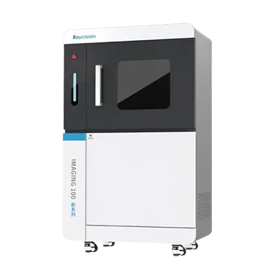
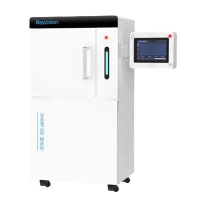
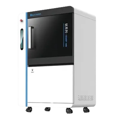
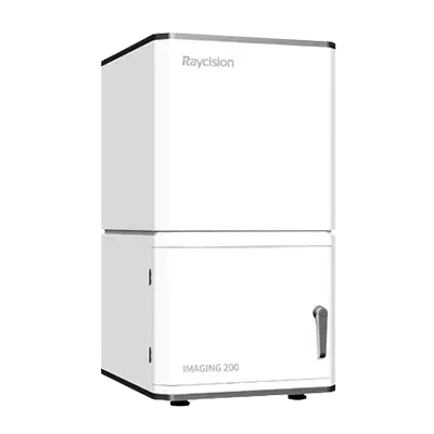
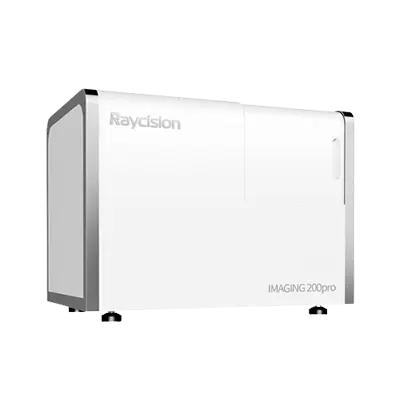
Raycision provides a range of preclinical imaging systems, including the IMAGING 100 In Vivo/Ex Vivo Micro-CT Imaging System, IMAGING 100pro Ultra-High-Resolution In Vivo/Ex Vivo Micro-CT Imaging System, IMAGING 200 2D Optical Molecular Imaging System, IMAGING 200pro 3D Optical Molecular Imaging System, and IMAGING 1000 Multimodality Precision Imaging System.
Traditional endpoint analysis provides detailed information at a specific experimental stage, but it cannot continuously show how a disease or treatment changes within the same animal. In vivo imaging enables repeated measurements at selected time points, allowing researchers to compare biological and anatomical changes throughout a study.
This longitudinal approach can support investigations into:
Tumor growth and metastasis
Drug distribution and treatment response
Bone development and remodeling
Cardiovascular structure and function
Infectious and inflammatory processes
Gene expression and molecular targets
Repeated in vivo animal imaging can also reduce variability caused by comparing different animals at every time point. Each animal can serve as its own reference, helping research teams observe individual changes more clearly.
Raycision imaging systems support research involving mice, rats, and other small-animal models, with technologies ranging from anatomical Micro-CT to 2D and 3D optical molecular imaging.
Micro-CT and optical molecular imaging provide complementary information, so the appropriate choice depends on the research objective.
Micro-CT uses X-ray projection data to reconstruct three-dimensional anatomical structures. It is particularly useful for examining bone morphology, lung architecture, organs, implants, vasculature, and other structures distinguishable by X-ray attenuation.
Raycision's IMAGING 100 is designed for both in vivo and ex vivo Micro-CT studies. It provides multiple fields of view, GPU-accelerated reconstruction, a minimum listed voxel size of 1 μm, and a spatial resolution of 5 μm. This enables one platform to support live-animal scanning as well as detailed imaging of isolated tissues or material samples.
For studies requiring ultra-high-resolution ex vivo imaging and fine microstructural analysis, Raycision's IMAGING 100pro provides a more specialized option. Designed as an ultra-high-resolution in vivo and ex vivo Micro-CT system, it delivers spatial resolution up to 3.6 μm and supports detailed visualization of structures such as trabecular bone, dental enamel, microvasculature, and implant interfaces.
Because it supports both in vivo and ex vivo imaging at high spatial resolution, the IMAGING 100pro is particularly useful when a study progresses from longitudinal in vivo observation to detailed ex vivo analysis. Researchers can monitor disease or anatomical changes in living animals and then examine excised tissues at higher structural detail after the selected experimental endpoint.
In addition to its resolution capabilities, the IMAGING 100pro also supports large-field-of-view imaging, high-precision gating, rapid volumetric reconstruction, and quantitative analysis for applications involving bone, lung, cardiovascular, vascular, and adipose tissue research.
Note:Voxel size refers to the smallest volume element in a reconstructed image, whereas spatial resolution describes the system’s ability to distinguish adjacent structures. These are different technical metrics and should not be compared solely by numerical value. Spatial resolution is a more direct indicator of the system’s resolving capability.
While Micro-CT excels at anatomical and structural imaging, it provides limited molecular information. Small animal optical imaging, in contrast, is designed to detect molecular and cellular signals with high sensitivity. Bioluminescence imaging can track reporter-expressing cells or biological activity, while fluorescence imaging can identify selected molecular targets.
The IMAGING 200 provides 2D optical molecular imaging with high-throughput operation, autofluorescence correction, wavelength-specific fluorescence excitation, and quantitative analysis. It is suitable when researchers need sensitive optical signal detection without full three-dimensional localization.
In summary, Micro-CT is the preferred choice when anatomical and structural information is the primary endpoint, while optical molecular imaging is more appropriate when molecular or cellular signals are the focus. When both are required, multimodality imaging may be considered.
Two-dimensional optical imaging can show signal intensity and approximate position, but depth and anatomical location may remain difficult to determine. For studies requiring more precise spatial information, three-dimensional optical imaging can provide greater value.
The IMAGING 200pro integrates bioluminescence tomography and fluorescence molecular tomography. Its reconstruction methods locate and quantify optical signals within three-dimensional space rather than only displaying their projection on the animal surface.
This capability may be useful for:
Localizing deep tumors or metastatic lesions
Comparing signal distribution between organs
Measuring changes in target volume
Studying the depth of fluorescent or bioluminescent sources
Improving longitudinal quantification
The choice between 2D and 3D optical imaging therefore depends on the study endpoint. High-throughput screening may favor 2D imaging, while small, deep, or spatially complex targets may require three-dimensional reconstruction.
No single imaging modality provides every type of biological information. Optical imaging offers high sensitivity to molecular signals, while Micro-CT provides detailed anatomical structure. Combining both can reveal not only whether a molecular target is present, but also where it is located relative to surrounding tissues.
Raycision's IMAGING 1000 integrates Micro-CT, bioluminescence imaging, and fluorescence molecular imaging in one platform. Its image-registration workflow coregisters reconstructed optical signals with Micro-CT data for three-dimensional visualization, localization, and quantitative analysis.
The system also supports GPU-accelerated CT reconstruction, bioluminescence tomography, fluorescence molecular tomography, multimodality image registration, 3D visualization, target quantification, and AI-powered organ contouring.
This approach can be particularly valuable in oncology research. Optical imaging may identify small tumor signals, while CT provides the anatomical framework needed to determine whether those targets are located in the liver, bone, lung, or other tissues.
Selecting an in vivo imaging system for small animals should begin with the biological question rather than a preferred technology. Research teams should define what they need to detect, whether animals will be scanned repeatedly, whether ex vivo analysis is required, and whether anatomical or molecular information is the primary endpoint.
| Selection factor | Key question |
|---|---|
| Imaging objective | Is the study focused on anatomy, molecular activity, or both? |
| Sample type | Will imaging involve living animals, ex vivo samples, or both? |
| Resolution | Is routine anatomical imaging sufficient, or is ultra-high-resolution analysis required? |
| Target depth | Are molecular signals superficial or deep within the animal? |
| Spatial information | Is 2D screening sufficient, or is 3D localization required? |
| Throughput | How many animals must be imaged per session? |
| Longitudinal design | How frequently will repeated imaging be performed? |
| Quantification | Are signal intensity, target volume, bone, or vascular measurements required? |
For flexible in vivo and ex vivo anatomical Micro-CT, IMAGING 100 provides multiple fields of view and rapid reconstruction. When studies require particularly detailed analysis of fine microstructures, IMAGING 100pro adds ultra-high-resolution in vivo and ex vivo Micro-CT capability, making it suitable for applications involving trabecular bone, microvasculature, dental structures, and implant interfaces.
Researchers should also compare software workflows, animal positioning, reconstruction speed, quantitative tools, gating capabilities, training, and technical support when selecting a system.
It is a research platform used to visualize anatomical structures or biological processes inside a living animal without immediately requiring tissue removal or endpoint analysis.
In vivo imaging examines a living animal, while ex vivo imaging analyzes tissues, organs, or samples outside the living organism. Raycision's IMAGING 100 and IMAGING 100pro both support in vivo and ex vivo Micro-CT workflows.
The IMAGING 100pro is designed for ultra-high-resolution in vivo and ex vivo Micro-CT. Its spatial resolution reaches up to 3.6 μm, supporting detailed visualization of fine structures such as trabecular bone, dental enamel, microvasculature, and implant interfaces.
Yes. Bioluminescence and fluorescence imaging can be repeated at planned intervals to monitor molecular changes, disease progression, or treatment response.
Multimodality imaging is useful when molecular signals need to be accurately related to anatomical structures or quantified within three-dimensional space.
The IMAGING 200pro provides 3D optical molecular imaging, while the IMAGING 1000 combines 3D optical imaging with Micro-CT in a multimodality platform.
In vivo imaging systems allow researchers to observe anatomical, molecular, and functional changes throughout preclinical studies. Micro-CT supports structural analysis, optical molecular imaging provides sensitive detection of biological targets, and multimodality imaging combines molecular and anatomical information for more precise localization.
For studies requiring detailed analysis after longitudinal animal imaging, the IMAGING 100pro extends Raycision's Micro-CT portfolio with ultra-high-resolution in vivo and ex vivo imaging, reaching spatial resolution up to 3.6 μm for fine structural investigations.
Raycision's portfolio therefore covers flexible Micro-CT with IMAGING 100, ultra-high-resolution Micro-CT with IMAGING 100pro, 2D optical imaging with IMAGING 200, 3D optical imaging with IMAGING 200pro, and integrated multimodality imaging with IMAGING 1000, helping laboratories select a platform aligned with both current research needs and future applications.
This is the first one.
 English